Feature Extraction and Matching
4D (Spatial-temporal) Volumetric Registration
Modeling Deformation of Lung
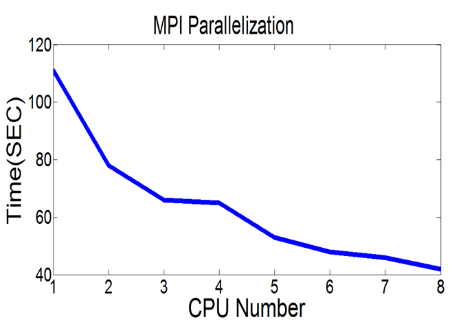
Parallel Speed-up (GPU/High-performance Computing)
Feature Extraction and Matching |
4D (Spatial-temporal) Volumetric Registration |
|
|
|
Modeling Deformation of Lung |
Parallel Speed-up (GPU/High-performance Computing) |
|
|
|
 |
1. H. Xu, Xin
Li, " Consistent Feature-aligned 4D Image Registration for Respiratory Motion Modeling.
," IEEE International Symposium on
Biomedical Imaging (ISBI) 2013 (Oral Paper) , [PDF]
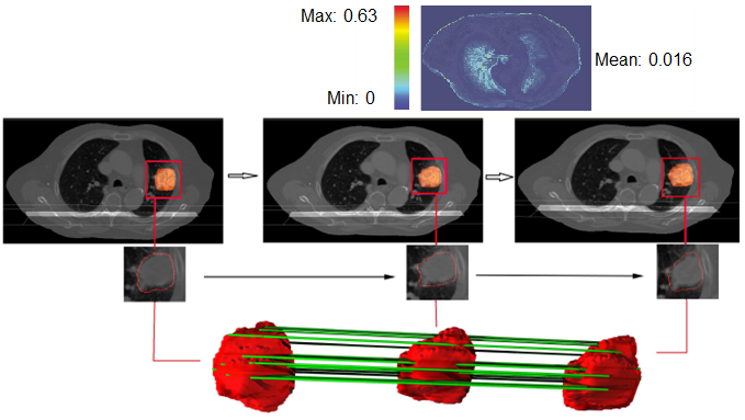
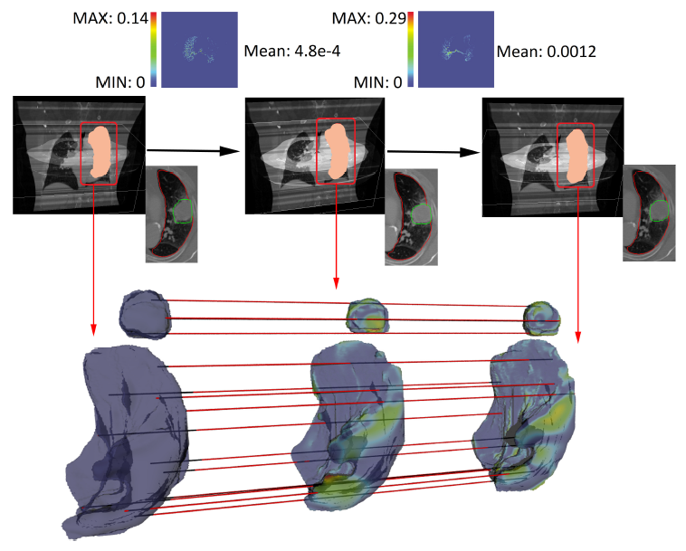
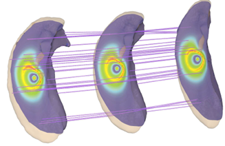
This paper presents a consistent feature-aligned 4D image registration algorithm and its medical application. The matching across a temporal sequence of volumetric images is based on a 4D (3D spatial + 1D temporal) free-form B-spline deformation model, which ensures interpolated motions with both spatial and temporal smoothness. We first develop the forward and inverse matching models with feature alignment constraints, then iteratively refine the registration results by incorporating extra inverse consistency. Experimental results show that our method achieves better registration accuracy than previous 3D registration and 4D registration methods. This algorithm can be used to parameterize temporal CT lung volume images for motion analysis and tracking. |
 |
2. S.
Iyengar, Xin Li, Huanhuan Xu, Amit Sawant, P. Iyengar, S. Mukhopadhyay, N.
Balakrishnan. "Toward More Precise Radiotherapy Treatment of Lung
Tumors," IEEE Computer, vol.45, no.1, pp.59-65, 2012. [PDF] [Bibtex]
A computational framework for modeling the respiratory motion of lung tumors provides a 4D parametric representation that tracks, analyzes, and models movement to provide more accurate guidance in the planning and delivery of lung tumor radiothearpy. |
| |
3. H. Xu, P. Chen, W.
Yu, A. Sawant, S. Iyengar, Xin
Li, " Feature-aligned 4D Spatiotemporal Image Registration.
," International Conference on
Pattern Recognition (ICPR) 2012 (Oral Paper), pages 2639-2642 , [PDF]
[Talk Slides]
[Video]
[Bibtex]
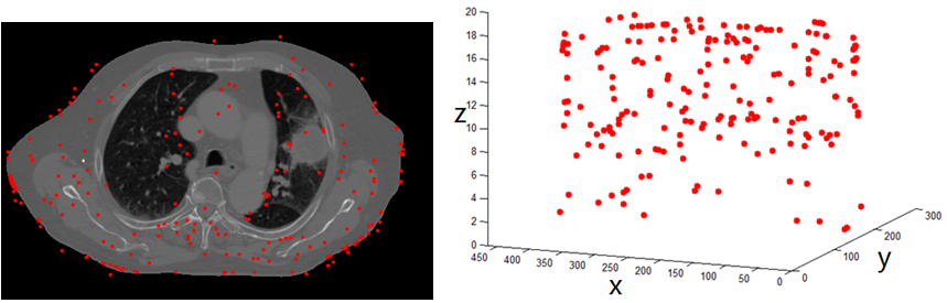
We develop a feature-aware 4D spatiotemporal image registration method. Our model is based on a 4D (3D+time) free-form B-spline deformation model which has both spatial and temporal smoothness. We first introduce an automatic 3D feature extraction and matching method based on an improved 3D SIFT descriptor, which is scale- and rotation- invariant. Then we use the results of feature correspondence to guide an intensity-based deformable image registration. Experimental results show that our method can lead to smooth temporal registration with good matching accuracy; therefore this registration model is potentially suitable for dynamic tumor tracking. |
 |
4. H. Xu, W. Yu, S.
Gu, Xin Li,
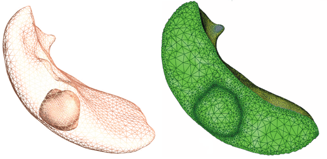
Biharmonic Volumetric Mapping using Fundamental Solutions ,
IEEE Trans. on Visualization and Computer
Graphics, in press, 2013.
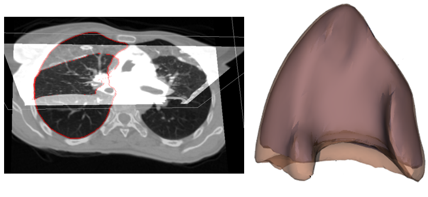
We propose a biharmonic model for cross-object volumetric mapping. This new computational model aims to facilitate the mapping of solid models with complicated geometry or heterogeneous inner structure. We demonstrate its effectiveness on the temporally scanned lung data collected during multiple respiratory cycles of some patient having the lung tumor. [Paper][Bibtex] |
Feature-aligned Harmonic Volumetric Mapping using MFS: Remeshing David Head |