|
|
MEMS-based optical probes for endoscopic optical coherence tomography (EOCT)
OCT is analogous to ultrasonography. Figure 1 shows the schematic block diagram of an OCT system.
- OCT uses light (typically near-infrared or NIR) instead of acoustic wave.
- Axial focusing is achieved by interference of short-coherent-length light.
- Axial scanning is obtained by scanning optical delay.
- OCT is useful as an in vivo imaging tool, when combined with endoscopy.
- There are two types of configurations depending on the imaging orientation of the probe relative to its guiding direction.
- Forward-looking probes are suitable for hollow organs.
- Side-looking probes are for luminal structures.
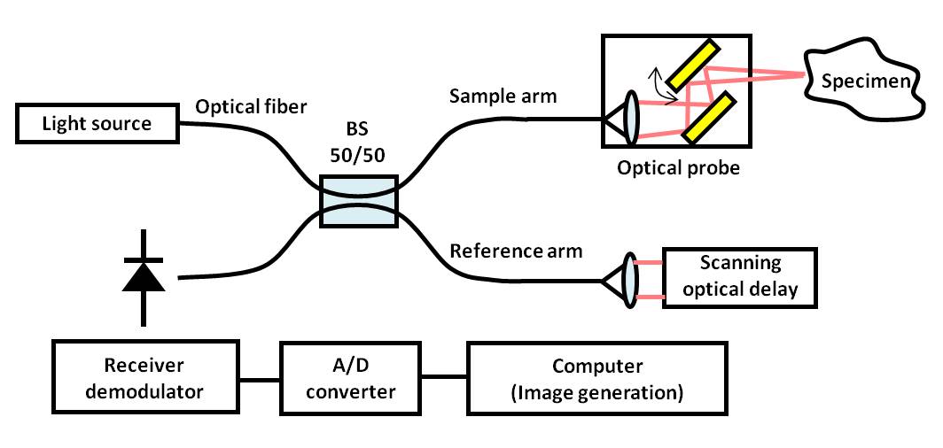
Fig. 1. Schematic block diagram of a typical OCT system.
MEMS microscanner for transversal scanning
- There are various ways to implement transversal scanning in OCT.
- Mechanical rotation of probe with external motor
- Scanning a mirror with an internal galvanometric motor
- Fiber tip swing with a piezoelectric cantilever
- Proximal scanning with relay lenses
- Fused multimode fiber bundles
- Grin rod lens array
- MEMS microscanners bring following advantages.
- Compact probe
- Minimal vibration
- No intermediate mechanical devices necessary, such as links
A narrow, forward-looking optical probe (Figure 2)
- Light passes a GRIN lens three times for one direction.
- Scanning mirror reflects the light almost parallel to the optical axis.
- Light stays more or less parallel to the optical axis, resulting in small probe diameter.
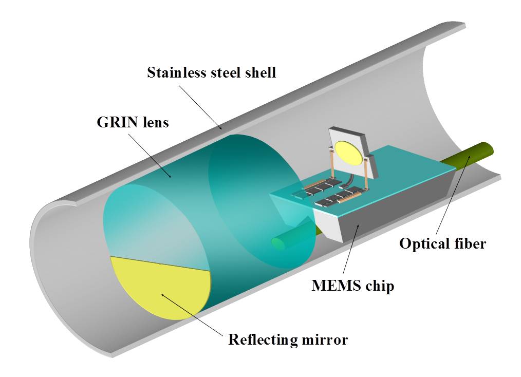
Fig. 2. Schematic diagram of the narrow, forward-looking optical probe.
|
